The sequencing of the entire human genome
back in the early 2000s is considered one of the most important scientific
breakthrough enabling data-driven medicine.
It flooded researchers in biology with a
tremendous amount of Big Data that needed to be processed and tested. It was
around then that DNA microarrays, small glass chips where thousands of DNA
fragments are deposited, became popular because they enable high-throughput and
multiplexed testing for gene expression and mutations, for example in cancer.
 |
| From left to right, Julien Cors, Govind Kaigala and David Taylor |
Interestingly, DNA and protein microarrays were one
of the convergence points of microtechnology and biology that resulted in the
fabrication of high-density arrays of biomolecules.
While protein microarrays have significant value for drug discovery, and
molecular profiling, the same success in the use of DNA arrays was not
transferable to protein arrays due to the limited quality of protein arrays.
This inspired a team of IBM scientists
to develop a new biopatterning method for efficient, accurate and high-quality
patterning of proteins on surfaces by addressing fundamental bottlenecks
inherent to the fabrication of protein microarrays. Such high-quality
microarrays will benefit quantitative biological assays for personalized
diagnostics and screening applications.
A new paper appearing this month on the
cover of Analytical
Chemistry details their latest research breakthrough. I spoke with two
of the authors Govind
Kaigala and Julien
Cors.
 |
Cover of Analytical Chemistry
Credit: ACS Publications |
One of the benefits of your technique is depositing
reagents on surfaces in a wet environment. Why is this important and how does
it compare to current approaches?
Govind Kaigala (GK): Current techniques for protein patterning use
inkjet and pin spotters, and they do not produce homogeneous protein patterns
-- largely because they operate in a dry environment.
So we asked ourselves, biology is wet, so how
can we accurately test samples when we are changing it’s make up by drying it
out?
In contrast, our method relies on a microfluidic
probe (MFP) that confines nanoliter volumes of reagents in a wet
environment. By fully immersing the substrate in a liquid, the “coffee
stain effect” (Marangoni
effect), is minimized with the reduction of evaporation. In
addition, we use continuous flows on the surface, which ensure improved reaction
kinetics compared to deposition relying on diffusion in existing techniques.
Also via a technique of re-circulating liquid we improve
reagent utilisation, which is important because its very expensive.
In our article we studied the
transport of biomolecules in flows and their surface interactions and to
address this we designed specific MFP heads to create homogeneous protein
patterns with volume consumption comparable to inkjet spotters.
The paper also describes the
convection-enhanced transport and recirculation of sub-microliter volumes using
analytical models.
A major factor of your research is working at the
micrometer level. Can you explain what the trick is when working with such
small sample sizes?
Julien Cors (JC): IBM is considered the birthplace for
scanning probe techniques with the original scanning probes developed for
atomic imaging. Our scanning probe isn’t imaging atoms, the MFP technology that
we are developing is for biological applications, in this case protein
patterning. But, the technique is similar in that the probe head never comes
into contact with the sample and is used in liquid environments and with a
resolution of a few micrometers.
By continuously injecting and
aspirating liquids, nanoliter volumes of liquids are confined at the tip of the
head. To put this into perspective, we are working with volumes of liquids
thousands of times smaller than a tear drop.
At this micrometer-length scale both reaction
kinetics and transport of biomolecules onto the surface are
highly favourable By exposing one or multiple such confined liquid
using the probe on a surface diverse patterns of biomolecules can be created.
Can you talk about some specific applications for
the innovation?
GK: Protein-based assays
(immunoassays) are very broadly applicable, and our method can be used to
create high-quality substrates to implement such tests.
Current protein microarrays provide
qualitative information i.e. presence or absence of a specific biomarker.
Because of the inhomogeneity of the deposited patterns, extracting quantitative
information remains challenging. Quantitative data have great importance both
in diagnostics and biomarker discovery.
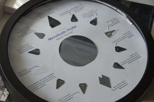 |
The MFP comes with different microfluidic
patterns based on the type of application. |
For example, in the Analytical Chemistry paper we demonstrate an
assay for the detection immunoglobulins (IgGs). IgGs are used as a diagnostics
marker for several autoimmune diseases and as a measure of the immune response.
This
research is partially funded by your European Research Council (ERC) grant.
What do you hope to achieve by the end of the grant?
GK: Within the ERC-BioProbe project, we
are developing new concepts, tools and methods for improved molecular profiling
of tissues for tumor diagnosis for personalized medicine.
Techniques for liquid recirculation and analytical models for
improved protein microarrays was in fact an off-shoot of this work – while in
BioProbe, we will focus on the main theme, we keep an eye for such projects and
pursue them.
What's
next for your research and when can we expect it to be used outside of IBM by
partners?
GK: We are very excited about the progress and evolution of the
microfluidic probe, particularly as it is now starting to be applied in various
areas including pathology and personalized medicine.
Labels: healthcare, IBM Research - Zurich, microfluidic probe

